It is a new generation product and is the follow-up model of EPIPHOT TME300/200 metallographic microscope. It supports bright field, dark field, differential interference, fluorescence, simple polarization and other observation methods. It has compact structure, convenient operation, uniform illumination, and clear imaging. , Energy-saving, power-saving, durable and other features.

Easy to operate
The parts that often need to be manipulated are concentrated near the observer, that is, in front of the microscope. Such as: field diaphragm, aperture diaphragm, polarizer, analyzer, inspection board insertion and removal, and switching between bright and dark fields.

Compact structure, small footprint, vibration resistance, sturdiness and durability
The box-shaped structure is firm and durable; compared with TME300, it has a smaller footprint and can save about 1/3 of the space.

High optical performance, uniform lighting, energy saving and power saving
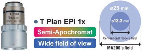
Adopting Nikon's unique CFI₆₀ optical system, not only can get a high contrast, clear bright field image, but also a dark field image with 3 times the brightness of the past; newly developed 1x and 40x objective lenses; eyepiece view The field number is up to 25mm, combined with the newly developed 1x objective lens, samples with a diameter of 25mm can be seen all at once; the illumination is uniform and the image is clear; the newly developed 12V50W halogen lamp illumination, its illumination brightness is completely comparable In the past, 12V100W halogen lamp lighting, and the electricity can be saved by half.
Photomicrography and image processing
Through the optional Nikon DS series digital camera, photomicrography can be carried out; the microscopic pictures taken can be observed and processed in two ways, respectively:

DS-L2
1. DS-L2 independent display controller (image data can be saved to U disk)

DS-U2
2. DS-U2 type computer is connected to the controller, the latter needs to be used with NIS Elements software
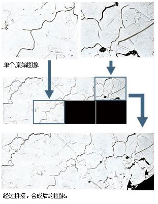
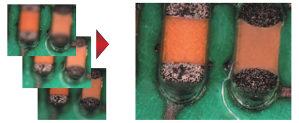
Use the functions in the NIS Elements software to realize seamless splicing of microscopic images;
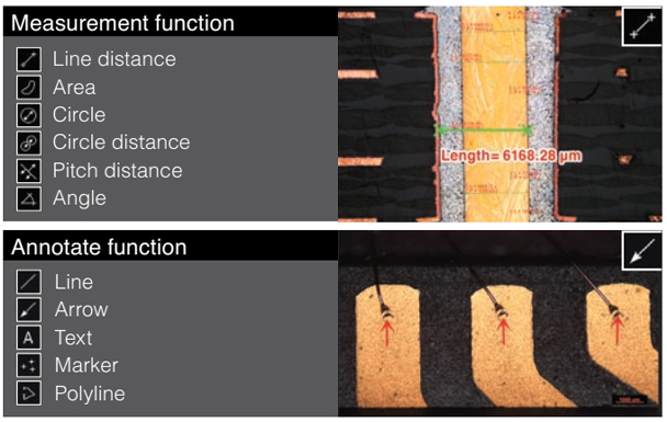
Using the functions in the NIS Elements software, you can perform material analysis, such as grain size analysis and graphite spheroidization rate analysis of ductile iron.
Offers high stability, durability, and a smaller footprint than conventional models, as well as easy access to the stage handle, the nosepiece, the BF/DF change lever, and diaphragms, all located on the front side.
| Compatible observation methods |
Δ: only available with Halogen Lamp and Fiber Illumination |
||||||||||||
|---|---|---|---|---|---|---|---|---|---|---|---|---|---|
| Compatible illminators |
|
||||||||||||
| Magnification module |
|
||||||||||||
| Compatible stages |
|
||||||||||||
| MA200 | ||
|---|---|---|
| Main body | Focusing mechanism | Focusing nosepiece (Fixed stage) Coaxial coarse/fine adjustment knob (torque adjustable) |
| Coarse adjustment of 4.0 mm per rotation, fine adjustment of 0.1 mm per rotation | ||
| Illumination | With flare prevention, Built in UV cut filter | |
| Field diaphragm: dialing continuous variable (centerable), Aperture diaphragm: dialing continuous variable (centerable) | ||
| Filter: Double turret (ND16, ND4/GIF, NCB, Additional option available), Polarizing block (Selectable with or without 1/4 Plate) | ||
| Fluorescence filter blocks: B/G/V/BV | ||
| 12V50W Halogen Lamp, C-HGFI HG Fiber Illuminator, LV-LL LED Lamphouse | ||
| Light distribution | Eyepiece tube/Back port: 100/0, 55/45 | |
| Optics | CFI₆₀/CFI₆₀-2 system | |
| Observation image | Surface Image | |
| Observation method | Bright/Darkfield/Simple Polarizing/DIC/Epi-Fluorescence | |
| Revolving nosepieces | LV-NU5I: Bright/Darkfield/DIC 5 position nosepiece, LV-NU5A: Motorized Bright/Darkfield/DIC 5 position nosepiece | |
| MA-N7-I Brightfield 7 position nosepiece (Intelligent) | ||
| Stage | MA2-SR Mechanical Stage (X/Y flexible handle) | |
| Dimension: 295×215 mm, Stroke: 50 mm×50 mm (with distance graduation), Standard accessory: ø22 universal specimen holder (with sample clip) | ||
| Trinocular eyepiece | Siedentopf interpupillary distance adjustment 50-75 mm | |
| Power source | 100-240 V, 50-60 Hz | |
| Power consumption (max.) | 1.2 A 75 W | |
| Weight | Approx. 26 kg (depends on combination) | |
| Options | Intermediate magnification | Turret (1×, 1.5×, 2×), Status detection (Output magnification information to main unit) |
| Scale | MA2-GR Grain Reticle (ASTM E112-63 grain sizing numbers 1 to 8), Grid Reticle(20 lines, 0.5 mm) | |
| MA2-MR Scale Reticle (compatible with 5-100×, Read in um, Dialing System) | ||
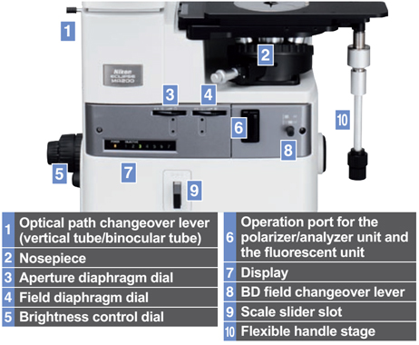 |
Front Operation Delivers ease-of-use by placing all important controls at the front of MA200N.
 The observation position of the objective lens and sample can be checked easily from the microscope’s front panel. |
| Evolved Optical Performance Provides a more ergonomic observation with clearer images.
Combine images with the stitching feature Can combine up to eight images with uniform lighting and no seams.  |
|
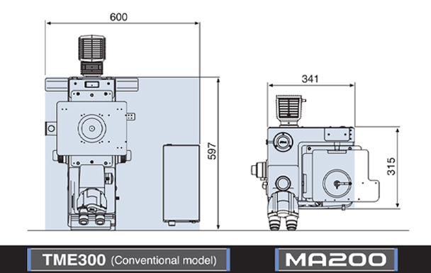 |
Box Structure The unique box structure is 1/3 smaller than conventional models and offers improved durability.
|
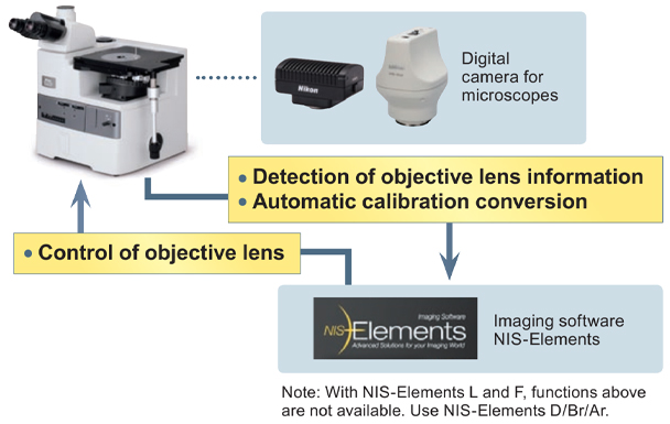 |
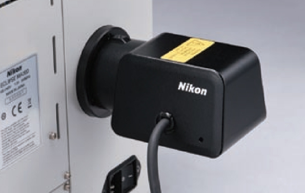
Combinaation with Digital Camera The MA200 allows detection of information and control of objective lenses, enabling optimization of the conditions vital for image acquisition. |
 LV-LL LED Lamphouse LV-LL LED Lamphouse |
Illumination Expanded lineup Added a compact LED illuminator to the existing lineup. With the use of LED, Nikon illuminators are power saving and achieve long life. |
Accessories Stage Samples can be rotated by the stage clip. The stage delivers high durability needed to support heavy samples.
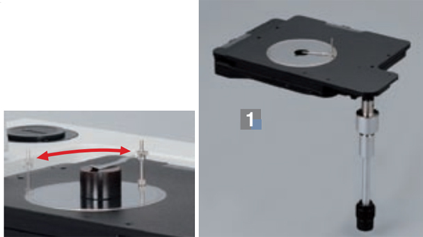 |
DIC Units Standard and high contrast type DIC prism are available to match needs of the sample. These prisms are effective for observation of minute step heights.
 |
 |
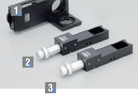
Nosepiece & Magnification Module Enables communication of objective lens position, magnification and intermediate magnification module information with the NIS-Elements image software.
|
Holders A full lineup is available that correspond to a variety of sample shapes. |
 |
Polarizing Units Polarizing observation is effective for birefringence samples. MA2-PA unit is suitable for observation of aluminium.
 |
 |
Grain Size Reticle & Scale Overlays a pattern onto the observed image. The Grain Size Reticle is used for grain size analysis and complies with the JIS G0551 and ASTM E112 standards. The Scale displays a scale for each objective lens magnification.
|
 |
Nikon's CFI₆₀ optical system, highly evaluated for its unique concept of high NA and long working distance, has achieved the apex in long working distance, chromatic aberration correction, and light weight.
Standard objective lenses
TU Plan Fluor Series
Enable brightfield, darkfield, simple polarizing, sensitive polarizing, differential interference, and epi-fluorescence observations with just one lens. Achieves superior chromatic aberration performance with long working distance for all magnifications to adapt to any application.

| Model | Magnification | NA | Working Distance(mm) |
|---|---|---|---|
| TU Plan Fluor EPI (brightfield type) |
5× | 0.15 | 23.5 |
| 10× | 0.30 | 17.5 | |
| 20× | 0.45 | 4.5 | |
| 50× | 0.80 | 1.0 | |
| 100× | 0.90 | 1.0 | |
| TU Plan Fluor BD (brightfield/ darkfield type) |
5× | 0.15 | 18.0 |
| 10× | 0.30 | 15.0 | |
| 20× | 0.45 | 4.5 | |
| 50× | 0.80 | 1.0 | |
| 100× | 0.90 | 1.0 |
Long working distance objective lenses
TU Plan ELWD Series
With the phase Fresnel lenses, these objective lenses enable long working distances while offering higher level chromatic aberration correction than conventional objective lenses. This improves operability for samples with different heights.

| Model | Magnification | NA | Working Distance(mm) |
|---|---|---|---|
| TU Plan EPI ELWD (brightfield type) |
20× | 0.4 | 19.0 |
| 50× | 0.6 | 11.0 | |
| 100× | 0.8 | 4.5 | |
| TU Plan BD ELWD (brightfield/ darkfield type) |
20× | 0.4 | 19.0 |
| 50× | 0.6 | 11.0 | |
| 100× | 0.8 | 4.5 |
Low-magnification objective lenses
T Plan EPI
Both clear observation using a conventional analyzer/polarizer and operability-oriented observation without the need of an analyzer/ polarizer are possible.

| Model | Magnification | NA | Working Distance(mm) |
|---|---|---|---|
| T Plan EPI (brightfield type) |
1× | 0.0.3 | 3.8 |
| 2.5× | 0.075 | 6.5 |
Apochromatic objective lenses
TU Plan Apo Series
By using phase Fresnel lenses, these objective lenses achieve significantly longer operating distances while maintaining the superior chromatic aberration performance of apochromatic lenses.

| Model | Magnification | NA | Working Distance(mm) |
|---|---|---|---|
| TU Plan Apo EPI (brightfield type) |
50× | 0.8 | 2.0 |
| 100× | 0.9 | 2.0 | |
| 150× | 0.9 | 1.5 | |
| TU Plan Apo EPI (brightfield/ darkfield type) |
50× | 0.8 | 2.0 |
| 100× | 0.9 | 2.0 | |
| 150× | 0.9 | 1.5 |
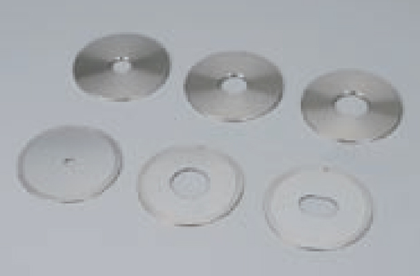
| Other Lenses Brightfield objective lense CFI L Plan EPI 40x A 40x objective lens is best for metal analysis. NA: 0.65 W.D.: 1.0 mm |
 |
Digital camera system for microscopes
DIGTAL SIGHT SERIES
| Microscope camera DS-Fi3 Three main features of the previous models, high-resolution, high sensitivity and low noise, and high-speed live display are offered in 1 camera. 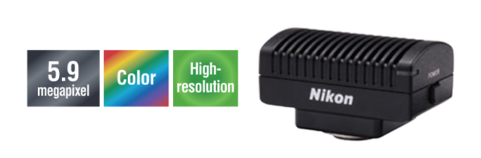 |
DS-Ri2 Capable of expressing images as is, this microscope digital camera offers high resolution, color reproduction, and frame rate. 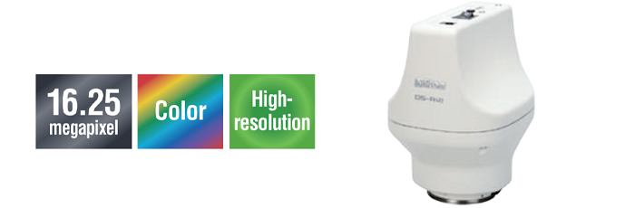 |
| Frame Rate | 30 fps (1440×1024) | 45 fps (1636×1088) |
|---|---|---|
| Max Recordable Pixels | 2880×2048 | 2880×2048 |
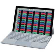
| Imaging software NIS-Elements Simply installing NIS-Elements L on a tablet PC enables setting and control of DS-Fi3/ DS-Ri2 microscope cameras, live image display, and image acquisition. |
 |
|
 |
|
 |
|
 |
|
 |
| * See the "Digital Camera Digital Sight Series for Microscopes" brochure for details on Digital Sight features. | |
System Diagram (MA200)


