Product Specification
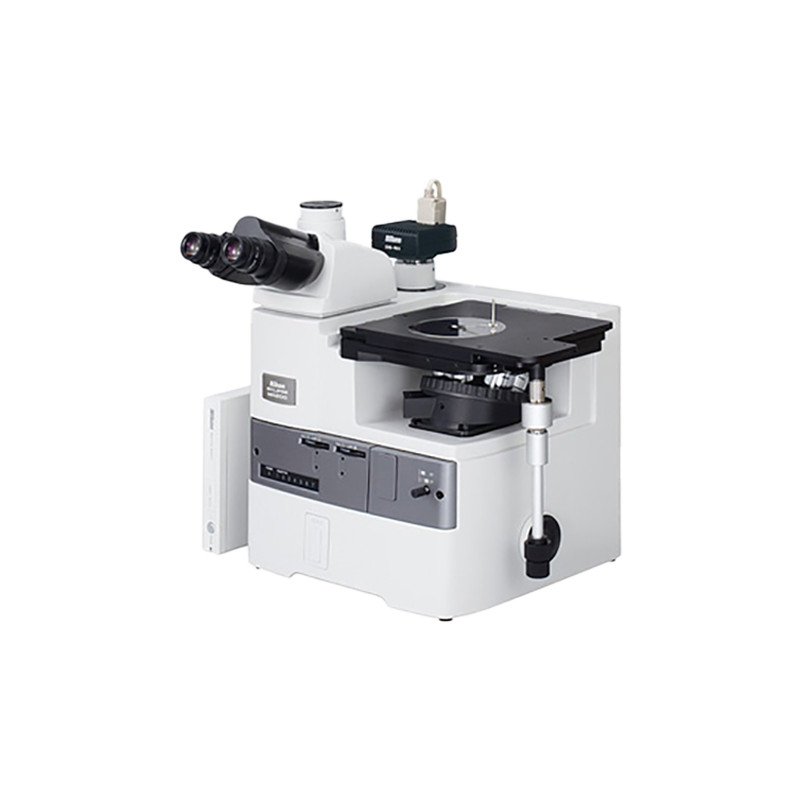
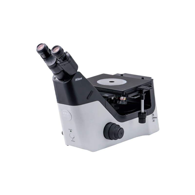
Designated for brightfield and simple polarizing observation, the MA100 is a cost-effective solution to manufacturing and QA/QC situations in industries such as automotive/electronic parts and industrial machinery/tools.
| Compatible observation methods |
|
||||||||||||
|---|---|---|---|---|---|---|---|---|---|---|---|---|---|
| Compatible illminators | High-intensity white LED Illuminator (internal power supply) | ||||||||||||
| Magnification module | - | ||||||||||||
| Compatible stages |
|
||||||||||||
| MA100N | |
|---|---|
| Optics | CFI₆₀/CFI₆₀-2 system |
| Observation image | Reversed image |
| Observation method | Brightfield and polarization (with MA P/A simple polarizer/analyzer set) |
| Focusing | Focusing nosepiece (fixed stage), coaxial coarse/fine adjustment knob with 8.5-mm stroke |
| (Coarse adjustment of 37.7 mm per rotation, fine adjustment of 0.2 mm per rotation) | |
| Nosepiece | Brightfield 5-position nosepiece |
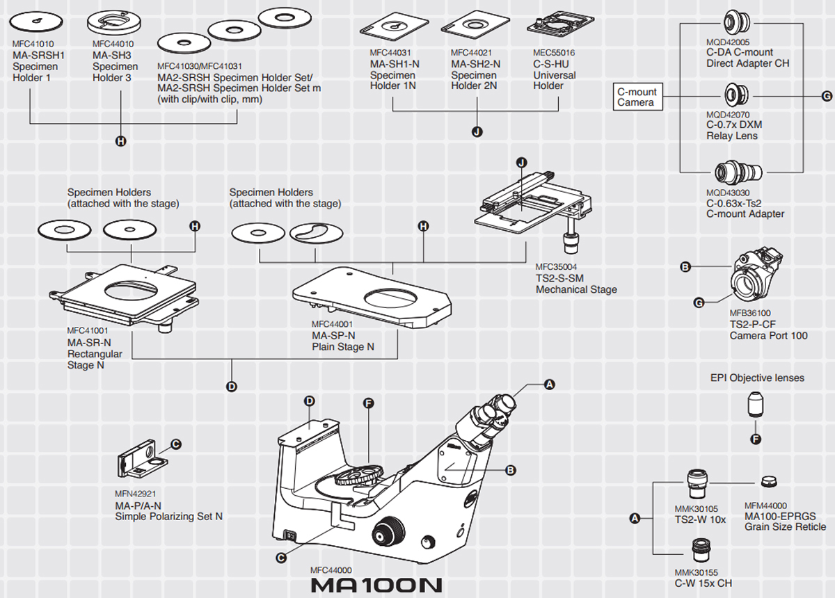
| Stage | MA-SR-N Rectangular 3-plate Stage N: 50×50 mm stroke (includes two stage inserts (ø20 mm and 40 mm opening) and coaxial control handle on the right side |
| The 3-plate design allows entire top surface to move. Optional Stage inserts: MA-SRSH1 Specimen Holder 1 with (ø15 mm opening or MA-SH3 | |
| Specimen Holder 3 with 2 mm to 32 mm adjustable opening | |
| MA-SP-N Plain Stage N: 188×310 mm - Includes two stage inserts (1) clear acrylic stage insert with ø30 mm opening, (2) clear acrylic stage insert | |
| with crescent opening (width 30 mm) to allow clearance for rotation of high magnification objectives | |
| Optional stage inserts: MA-SRSH1 Specimen Holder 1 with 15 mm opening or MA-SH3 Specimen Holder 3 with 2 mm to 32 mm adjustable opening | |
| Accepts Attachable Mechanical Stage TI-SM | |
| TS2-S-SM Mechanical Stage: 126 mm×78 mm stroke, handle can be attached on the right or left side of the plain stage | |
| Optional Specimen Holders to fit Attachable Mechanical stage: MA-SH1-N Specimen Holder 1N (ø15 mm opening) | |
| MA-SH2-N Specimen Holder 2N (ø30 mm opening), or C-S-HU Universal Holder (30 mm to 65 mm adjustable opening) | |
| Illuminator | Internal power supply white LED light source, condenser built-in (lever operated), ø25 mm filter can be inserted |
| Binocular body | Built-in Siedentopf binocular, 45 inclination angle and 50 to 75-mm interpupillary adjustment, attachable camera port, eyepiece/Port: 100/0:0/100 |
| Power consumption (max.) | 15W |
| Weight | Approx. 10 kg |
Illumination
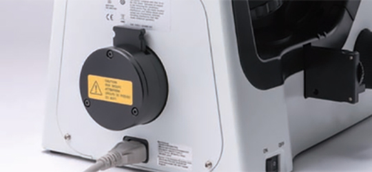
Employment of high-intensity LED illumination (Eco-illumination)
Compared to conventional halogen illumination, these high intensity LED sources need only about one third of consuming electricity and last approximately 30 times longer. The MA100N ensures stable sample observation with uniform color temperature even in different light intensity.
Stage
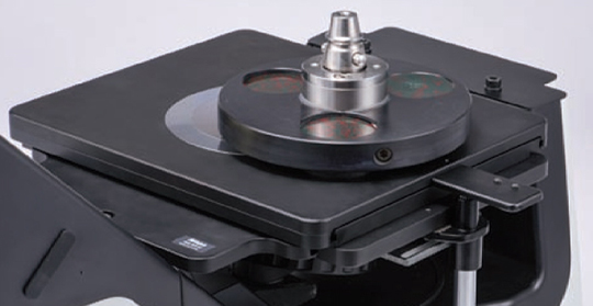
Controlled stability even with heavy samples/ Boasts superior durability
The MA-SR-N Rectangular Stage was developed especially for the MA100N. The three-plate structure allows for observation of heavy samples, such as a grinder resin mounted samples.
Compact Body
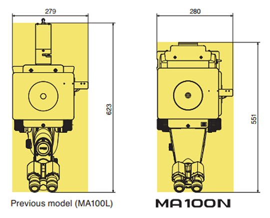
Redesigned to be smaller
Designed for LED illumination, the footprint is 11% smaller than conventional models, allowing users to have more installation choices.
Aperture Diaphragm
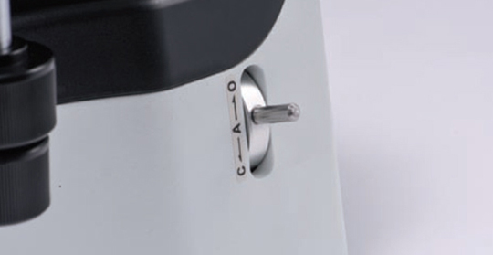
Standard with MA100N
The epi illuminator comes standard with a variable aperture diaphragm to control image contrast and depth of field.
Accessories
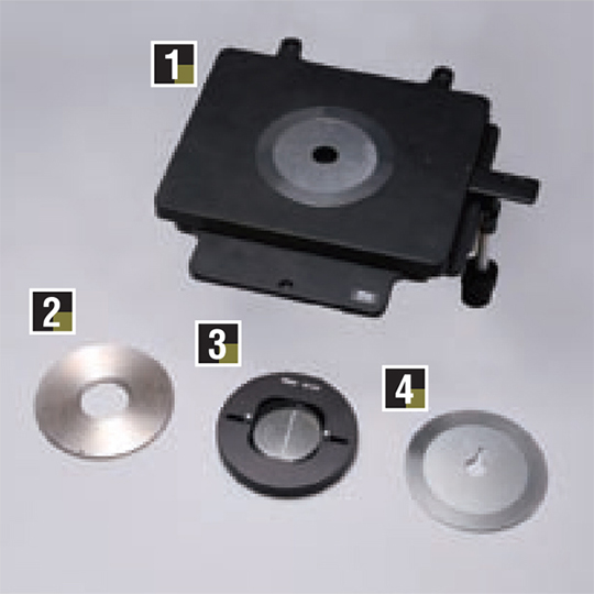
Basic stage set
A triple-platform stage structure lets you use heavy samples.
- MA-SR-N Rectangular Stage N
- Specimen Holder (ø20/30/40 mm aperture)
- MA-SH3 Specimen Holder 3
- MA-SRSH1 Universal Specimen Holder
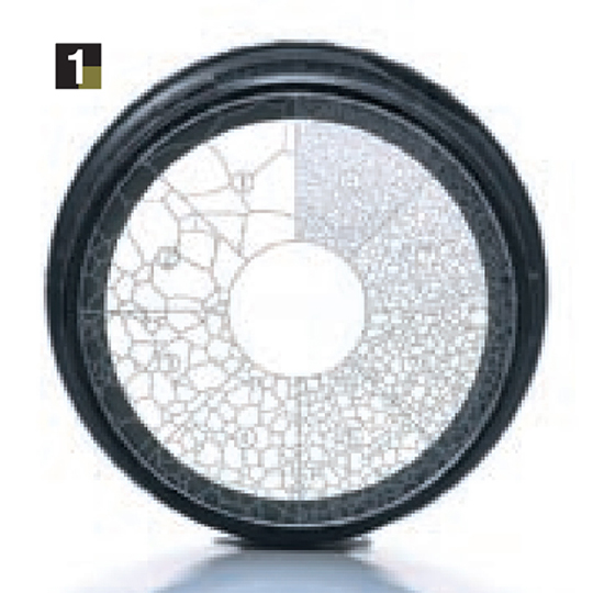
Grain size reticle
The class of grain size in a sample can be easily distinguished while observing its image.
1. MA100-EPRGS Grain Size Reticle
Digital Camera
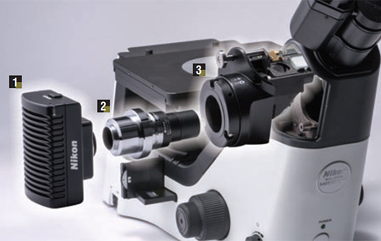
Redesigned with optical systems suitable for sample observations. The camera port is located on the side of MA100N to provide improved visibility of the stage.
- Microscope Camera DS-Fi3
- C-0.63x-TS2 C-mount Adapter
- TS2-P-CF Camera port 100
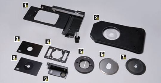
Other accessories
- TI-SM Mechanical Stage CH
- MA-SP-N Plain Stage N
- MA-SH2-N Specimen Holder 2N
- MA-S-HU Universal Holder
- MA-SH3 Specimen Holder 3
- MA-SRSH 25-40 Holder
- MA-SRSH1 Universal Specimen Holder
- MA-SH1-N Specimen Holder 1N
- MA-P/A Simple Polarizer
Nikon's CFI₆₀ optical system, highly evaluated for its unique concept of high NA and long working distance, has achieved the apex in long working distance, chromatic aberration correction, and light weight.
Standard objective lenses
TU Plan Fluor Series
EPI / BD|5x / 10x / 20x / 50x / 100x
Enable brightfield, darkfield, simple polarizing, sensitive polarizing, differential interference, and epi-fluorescence observations with just one lens. Achieves superior chromatic aberration performance with long working distance for all magnifications to adapt to any application.
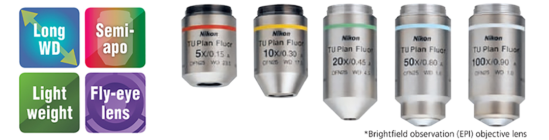
| Model | Magnification | NA | Working Distance(mm) |
|---|---|---|---|
| TU Plan Fluor EPI (brightfield type) |
5× | 0.15 | 23.5 |
| 10× | 0.30 | 17.5 | |
| 20× | 0.45 | 4.5 | |
| 50× | 0.80 | 1.0 | |
| 100× | 0.90 | 1.0 | |
| TU Plan Fluor BD (brightfield/ darkfield type) |
5× | 0.15 | 18.0 |
| 10× | 0.30 | 15.0 | |
| 20× | 0.45 | 4.5 | |
| 50× | 0.80 | 1.0 | |
| 100× | 0.90 | 1.0 |
Long working distance objective lenses
TU Plan ELWD Series
EPI / BD|20x / 50x / 100x
With the phase Fresnel lenses, these objective lenses enable long working distances while offering higher level chromatic aberration correction than conventional objective lenses. This improves operability for samples with different heights.
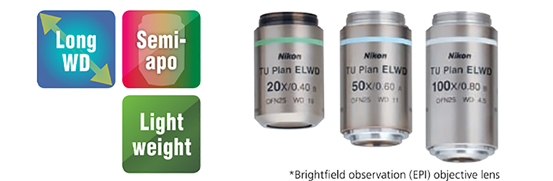
| Model | Magnification | NA | Working Distance(mm) |
|---|---|---|---|
| TU Plan EPI ELWD (brightfield type) |
20× | 0.4 | 19.0 |
| 50× | 0.6 | 11.0 | |
| 100× | 0.8 | 4.5 | |
| TU Plan BD ELWD (brightfield / darkfield type) |
20× | 0.4 | 19.0 |
| 50× | 0.6 | 11.0 | |
| 100× | 0.8 | 4.5 |
Low-magnification objective lenses
T Plan EPI
EPI|1x / 2.5x
Both clear observation using a conventional analyzer/polarizer and operability-oriented observation without the need of an analyzer/ polarizer are possible.
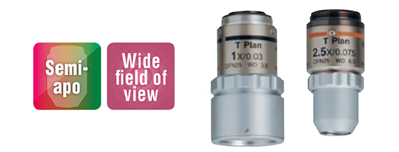
| Model | Magnification | NA | Working Distance(mm) |
|---|---|---|---|
| T Plan EPI (brightfield type) |
1× | 0.03 | 3.8 |
| 2.5× | 0.075 | 6.5 |
Apochromatic objective lenses
TU Plan Apo Series
EPI / BD|50x / 100x / 150x
By using phase Fresnel lenses, these objective lenses achieve significantly longer operating distances while maintaining the superior chromatic aberration performance of apochromatic lenses.
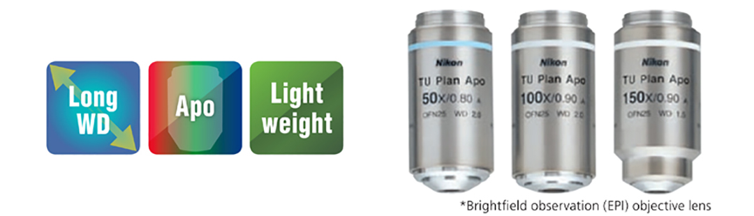
| Model | Magnification | NA | Working Distance(mm) |
|---|---|---|---|
| TU Plan Apo EPI (brightfield type) |
50× | 0.8 | 2.0 |
| 100× | 0.9 | 2.0 | |
| 150× | 0.9 | 1.5 | |
| TU Plan Apo EPI (brightfield/ darkfield type) |
50× | 0.8 | 2.0 |
| 100× | 0.9 | 2.0 | |
| 150× | 0.9 | 1.5 |
Other Lenses
Brightfield objective lense
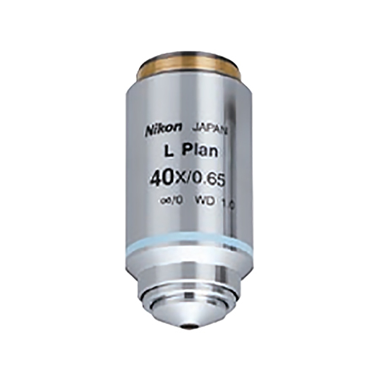
CFI L Plan EPI 40x
A 40x objective lens is best for metal analysis.
NA: 0.65 W.D.: 1.0 mm
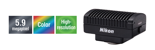
Digital camera system for microscopes
DIGTAL SIGHT SERIES
DS-Fi3 Microscope camera
Three main features of the previous models, high-resolution, high sensitivity and low noise, and high-speed live display are offered in 1 camera.
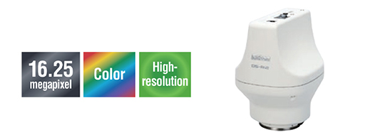
DS-Ri2
Capable of expressing images as is, this microscope digital camera offers high resolution, color reproduction, and frame rate.
| Frame Rate | 30 fps (1440×1024) | 45 fps (1636×1088) |
|---|---|---|
| Max Recordable Pixels | 2880×2048 | 2880×2048 |
Imaging software
NIS-Elements
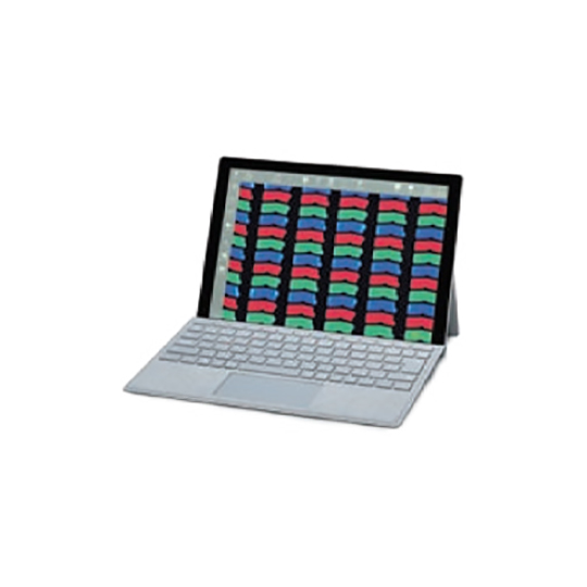
Using a tablet PC
Simply installing NIS-Elements L on a tablet PC enables setting and control of DS-Fi3/ DS-Ri2 microscope cameras, live image display, and image acquisition.
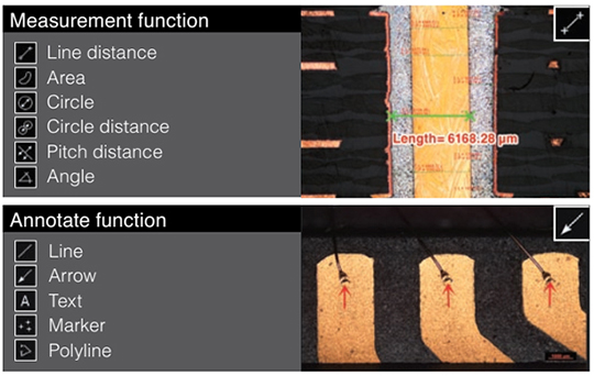
A wide variety of tools
NIS-Elements L enables the conducting of simple measurements on images, with input of lines and comments. These can also be written onto and saved with the image, and measurement data can be output.
Scene Mode
Ten camera setting patterns for optimal color reproduction and contrast for each microscope light source, observation method and type of sample, as well as custom settings, can be selected.
- Wafer / IC
- Metal, Ceramic / Plastic
- Circuit board
- Flat Panel Display
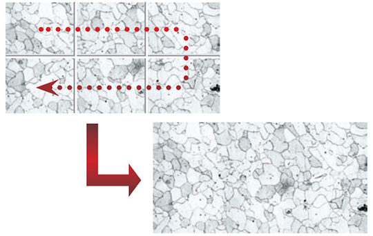
Image Stitching
Stitches together images acquired from multiple fields of view to create one image.
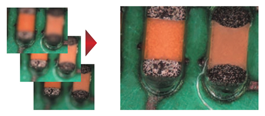
EDF (Extended Depth of Focus)
Create a single, all-in-focus image from images of differing focus.
* See the "Digital Camera Digital Sight Series for Microscopes" brochure for details on Digital Sight features.
System Diagram (MA100N)
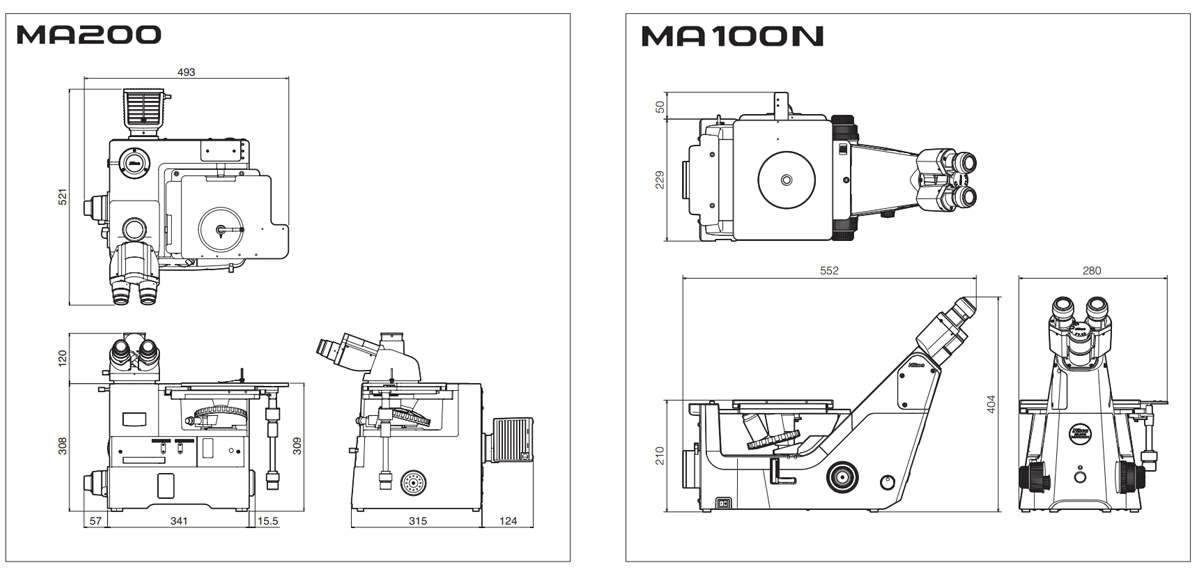
Dimensions